
Feline Leukaemia Virus (FeLV) is a retrovirus; a single stranded RNA virus belonging to the same viral family as the more common Feline Immunodeficiency Virus (FIV). It is found worldwide but incidence can be quite varied. In Australia, around 1-2% of the cat population is persistently infected with this virus, and many more are exposed. The proportion of cats infected differs according to the geographical location, environment and the lifestyle of the cat. For example, the prevalence in Western Australia (7-11%) is higher than that of the Eastern states (<2%). Infection is more common in colonies of cats where there is close contact between individuals and the virus is able to spread readily. The incidence of FeLV has dropped since the advent of effective vaccines and improved diagnostic techniques for the identification of infected cats.
Although many cats will overcome infection with FeLV, experiencing a transient viraemia or seroconverting with no detectable viraemia, some become persistently infected. Most persistently viraemic cats will develop a range of conditions including anaemia and/or lymphoma, cancers, intermittent immunosuppression and reproductive problems, and die within three years. In particular, cats may become susceptible to secondary infections if immunosuppressed. The cat’s age at the time of infection is a major determinant of clinical outcome. A cat found to be persistently viraemic should be isolated from other cats to reduce the risk of passing on the virus. A cat that has overcome viraemia will remain latently infected. Once a cat becomes latently infected, it remains so for life. FeLV may be reactivated on rare occasions in these cats when immunosuppressed or under chronic stress. Such cats should be considered potential sources of infection. Cats known to be persistently infected should be isolated from other cats to reduce the risk of passing on the virus.
FeLV is passed from cat to cat via saliva. Unlike FIV which is transmitted via biting, the transfer of FeLV is usually between friendly cats. This can occur during grooming or by sharing food bowls. Occasionally mothers can pass the infection to their kittens either in the womb or via milk. Kittens are particularly susceptible to contracting persistent infections, whereas most adult cats are able to eliminate the virus.
FeLV invades and replicates in cells of the cat’s immune system and blood-forming cells. During viral replication, the nucleic acid of FeLV inserts itself into the genome of the infected cells it has invaded. The result can be death of the cell or the viral insert being carried by the cell and passed on to the next generation during cell division. The change in the cell's genetic code can also potentially result in cellular changes that lead to neoplastic disease (cancer). The development of cancer or other conditions may not occur for months or years after the initial infection.
Polymerase chain reaction (PCR) detects the presence of infectious agents by identifying the genomic material of the agent being investigated. Unlike serology, which indicates whether an animal has been infected in the past, PCR determines if the agent is still present thereby informing the clinician that an active infection is in progress. It is often more sensitive and specific than other available tests including culture (especially for viruses) and is usually faster to achieve a result.
This test is a quantitative PCR test for the detection of FeLV proviral DNA. The FeLV PCR will detect the majority of strains of the virus.
Species:
Feline
Specimen:
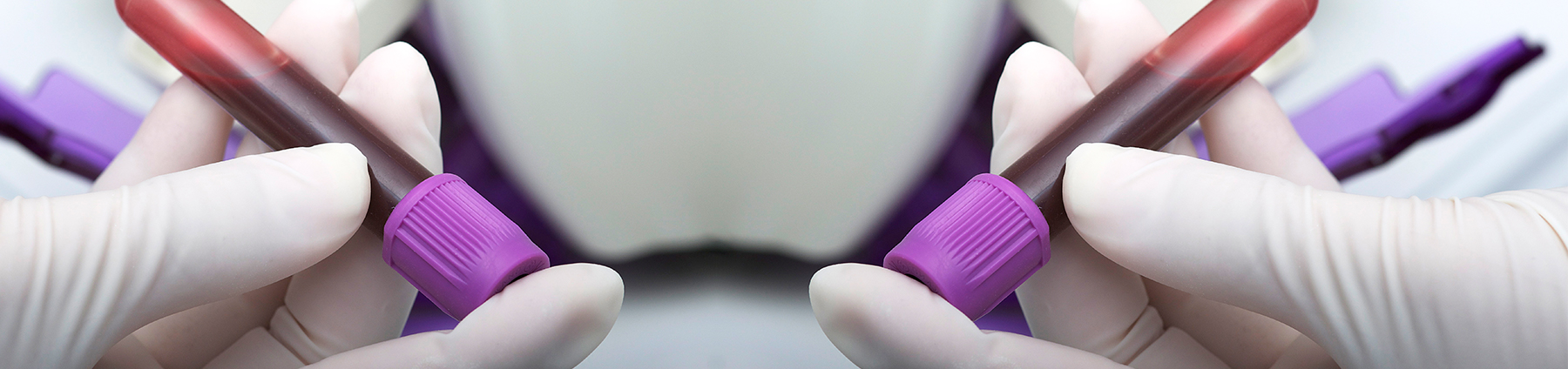
0.5-1 ml of whole blood, or bone marrow aspirate (0.5 ml in an EDTA tube)
Container:
EDTA, Lithium Heparin or Sodium Citrate
Collection protocol:
Standard venepuncture or bone marrow biopsy
Special handling/shipping requirements:
Blood samples are stable at room temperature and can be sent un-refrigerated. If collecting in warm conditions, it is recommended that samples be stored out of the heat, preferably in an insulated container, until they can be transferred indoors to a controlled environment.
References
- Lutz, H., Addie, D., Belák, S., Boucraut-Baralon, C., Egberink, H., Frymus, T., Gruffydd-Jones, T., Hartmann, K., Hosie, M.J., Lloret, A., Marsilio, F., Pennisi, M.G., Radford, A.D., Thiry, E., Truyen, U.and Horzinek, M.C. (2009). Feline leukaemia ABCD guidelines on prevention and management. Journal of Feline Medicine and Surgery 11 (7): 565-574.
- Malik, R. , Kendall, K. , Cridland, J. , Coulston, S. , Stuart, A.J. , Snow, D. and Love, D.N. (1997). Prevalences of feline leukaemia virus and feline immunodeficiency virus infections in cats in Sydney. Australian Veterinary Journal 75 (5): 323-327.
- Muirden, A. (2002). Prevalence of feline leukaemia virus and antibodies to feline immunodeficiency virus and feline coronavirus in stray cats sent to an RSPCA hospital. Veterinary Record 150 (20): 621-625.
- Tandon, R. , Cattori, V., Gomes-Keller, M.A., Meli, M.L., Golder, M.C., Lutz, H. and Hofmann-Lehmann, R. (2005). Quantitation of feline leukaemia virus viral and proviral loads by TaqMan® real-time polymerase chain reaction. Journal of Virological Methods 130 (1-2): 124-132.